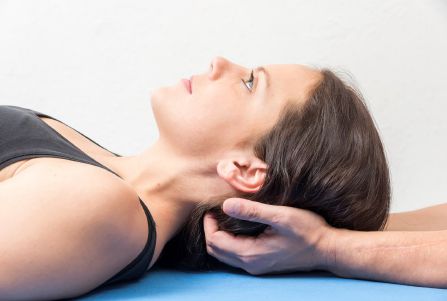
According to Roberts et al. (2021), end-diastolic velocity increased after performing occipitoatlantic decompression (OAD) (=technique for the atlantooccipital joint, Liem 2018, bilateral decoaptation of the atlantooccipital joint and decompression of the pars condylaris Liem 2020) in the middle cerebral artery (MCA), internal carotid artery (ICA), and vertebral artery (VA) (all p<0.001); no change occurred after sham contact (all p>0.05). This could be an explanation of how osteopathic manipulative treatment alleviates symptoms in patients with headache (Voigt et al 2011).
Objective: To investigate blood flow in the MCA, ICA, and VA before and after occipitoatlantic decompression (OAD) using Doppler sonography.
Methods: Thirty healthy osteopathic students (11 men, 19 women; mean age 24 years) in their first year of study at the Kirksville College of Osteopathic Medicine of A.T. Still University participated in a randomized, single-blinded crossover study with two treatments over two periods. Participants were randomly assigned to one of 2 treatment methods: OAD or sham contact. After one week, participants returned to receive the other treatment. Blood flow parameters—peak systolic velocity (PSV) and end-diastolic velocity (EDV)—in the middle cerebral artery (MCA), internal carotid artery (ICA), and vertebral artery (VA) were assessed before, immediately after, 5 minutes after, and 10 minutes after treatment. Differences in PSV, EDV, heart rate (HR), and blood pressure (BP) for both interventions were analyzed for the four time points using mixed effects models.
Results: EDV was greater at all post-treatment time points after OAD in the MCA, ICA, and VA than after sham contact (all p<0.001).
Conclusion: There was an increase in EDV in the large cranial arteries after OAD, but not after sham treatment. The exact mechanism of this increase is not known. Parasympathetic stimulation via secretion of vasodilating neurotransmitters or a decrease in external tissue pressure on the internal carotid artery (ICA) and on the vertebral artery (VA) are suspected, with the resulting flow causing further dilatation in the middle cerebral artery (MCA).
Roberts B, Makar AE, Canaan R, Pazdernik V, Kondrashova T. Effect of occipitoatlantal decompression on cerebral blood flow dynamics as evaluated by Doppler ultrasonography. J Osteopath Med. 2021 Feb 1;121(2):171-179. doi: 10.1515/jom-2020-0100.
https://pubmed.ncbi.nlm.nih.gov/33567080/
Voigt, K, Liebnitzky, J, Burmeister, U, et al.. Efficacy of osteopathic manipulative treatment of female patients with migraine: results of a randomized controlled trial. J Altern Complement Med. 2011;17(3):225-230.
Liem T. Praxis der Kraniosakralen Osteopathie, 2020, Thieme, Stuttgart.
Liem T. Kraniosakrale Osteopathie, 2018; Thieme, Stuttgart.
Occipitoatlantic Decompression Improves Blood Flow to the Brain
According to Roberts et al. (2021), after performing occipitoatlantic decompression (OAD) (=technique for the atlantooccipital joint, Liem 2018, bilateral decoaptation of the atlantooccipital joint and decompression of the pars condylaris Liem 2020), end-diastolic velocity increased in the middle cerebral artery (MCA), internal carotid artery (ICA) and vertebral artery (VA) (all p<0.001); no change occurred after sham contact (all p>0.05). This could be an explanation of how osteopathic manipulative treatment alleviates symptoms in patients with headache (Voigt et al 2011). Objective: To investigate blood flow in the MCA, ICA and VA before and after occipitoatlantic decompression (OAD) using Doppler sonography.Methods: Thirty healthy osteopathic students (11 men, 19 women; mean age 24 years) in their first year of study at the Kirksville College of Osteopathic Medicine of A.T. Still University participated in a randomised, single-blinded crossover study with two treatments over two periods. Participants were randomly assigned to one of 2 treatment methods: OAD or sham touch. After one week, participants returned to have the other treatment. Blood flow parameters – peak systolic velocity (PSV) and end-diastolic velocity (EDV) – in the middle cerebral artery (MCA), internal carotid artery (ICA) and vertebral artery (VA) were assessed before, immediately after, 5 minutes after and 10 minutes after treatment. Differences in PSV, EDV, heart rate (HR) and blood pressure (BP) for both interventions were analysed for the four time points using mixed effects models. Results: EDV was greater at all post-treatment time points after OAD in MCA, ICA and VA than after sham contact (all p<0.001).Conclusion: There was an increase in EDV in the large cranial arteries after OAD, but not after sham treatment. The exact mechanism of this increase is not known. Parasympathetic stimulation via secretion of vasodilating neurotransmitters or a decrease in external tissue pressure on the internal carotid artery (ICA) and on the vertebral artery (VA) are suspected, with the resulting flow causing further dilatation in the middle cerebral artery (MCA).
Roberts B, Makar AE, Canaan R, Pazdernik V, Kondrashova T. Effect of occipitoatlantal decompression on cerebral blood flow dynamics as evaluated by Doppler ultrasonography. J Osteopath Med. 2021 Feb 1;121(2):171-179. doi: 10.1515/jom-2020-0100.
https://pubmed.ncbi.nlm.nih.gov/33567080/
Voigt, K, Liebnitzky, J, Burmeister, U, et al.. Efficacy of osteopathic manipulative treatment of female patients with migraine: results of a randomized controlled trial. J Altern Complement Med. 2011;17(3):225-230.
Liem T. Praxis der Kraniosakralen Osteopathie, 2020, Thieme, Stuttgart.
Liem T. Kraniosakrale Osteopathie, 2018; Thieme, Stuttgart.