Bruxism and osteopathy
Summary
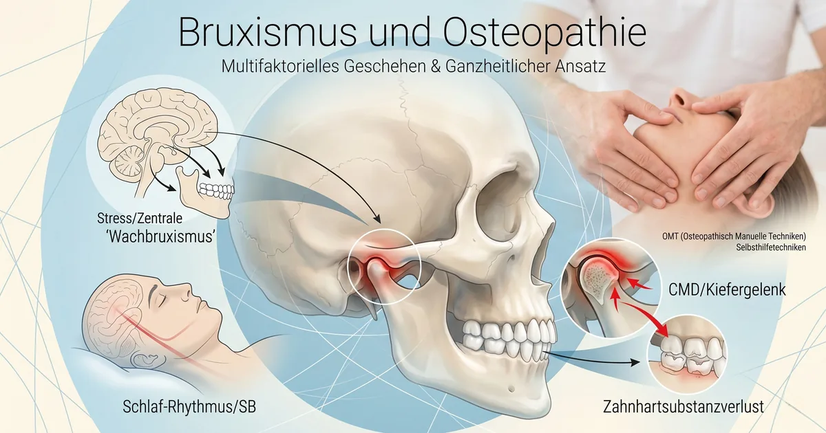
Bruxism can occur both as awake bruxism (WB) and as sleep bruxism (SB). The prevalence differs depending on the examination method and is given as 22.1-31% for WB and 12.8±3.1% for SB. The etiology of bruxism is only incompletely known, but it can be assumed to be multifactorial. The risk factors are presented in this article. Furthermore, neuroevolutionary and paleoanthropological perspectives are explained and insights into diagnostics are given. Finally, treatment approaches are described and the LEIT reset is presented, in which extrinsic (osteopathic manual techniques – OMT) and intrinsic jaw techniques (self-help techniques) are applied in a coordinated manner.
Keywords
Waking bruxism, sleeping bruxism, craniofacial pain, primary headache, temporomandibular joint disorder, teeth grinding, bruxism, dental hard tissue, manual therapy, craniomandibular dysfunction (CMD), osteopathic manual techniques, OMT, LEIT reset, self-help techniques
Summary
Bruxism can occur both as waking bruxism (WB) and as sleep bruxism (SB). The prevalence differs according to the method of investigation and is reported to be 22.1-31% in WB and 12.8±3.1% in SB. The etiology of bruxism is not completely known, but a multifactorial cause can be assumed. In this article, the risk factors are presented. Furthermore, neuroevolutionary and paleoanthropological perspectives are outlined and insights into diagnostics are given. (Manual therapy) treatment approaches and self-help techniques for patients are described. Finally, treatment approaches are described and the LEIT-Reset is presented – in which extrinsic (osteopathic manual techniques, OMT) and intrinsic jaw techniques (self-help techniques) are applied in a coordinated way.
Keywords
awake bruxism, sleep bruxism, craniofacial pain, primary headaches, temporomandibular disorder (TMD), tooth grinding, bruxism, hard dental tissue, osteopathic manual techniques (OMT), craniomandibular dysfunction (CMD), LEIT-Reset, self-help techniques.
A basic distinction is made between awake bruxism (WB) and sleep bruxism (SB). When awake, bruxism manifests itself through grinding or clenching of the teeth or spontaneous movements of the lower jaw without tooth contact [88]. According to the literature, these symptoms occur in around 20% of the adult population [47] and can lead to jaw pain, chewing muscle pain or headaches in both adults and children. A meta-analysis has shown that 1/6 of children and adolescents have clinical TMJ symptoms and over 23% of preschool children are affected by teeth clenching or grinding and pathological TMJ sounds [83]. Although the pathogenesis of TMJ is still unknown, stress, anxiety and nervous tics are considered risk factors [47].
Sleep bruxism is defined by the occurrence of the above-mentioned masticatory muscle activities during the sleep phase. A further distinction is made between rhythmic or phasic and non-rhythmic (short, repetitive contractions of the masticatory muscles with more than 3 muscle contractions of 0.25-2 sec) or tonic (muscle contractions >2 sec) activities. Furthermore, according to the ICD10 classification, SB does not represent a movement or sleep disorder in otherwise healthy people.
Bruxism can also be differentiated into primary (independent) and secondary (caused by other factors). Secondary bruxism can occur as a result of medication (antidepressants, anticonvulsants, antipsychotics, antihistamines, dopaminergic, cardioactive drugs) or drugs (especially smoking, alcohol, amphetamines, cocaine), but also as a comorbidity of certain diseases (especially coma and craniocerebral trauma) [88].
Relevance
While secondary bruxism can be treated, primary bruxism is currently considered incurable. This poses major challenges for various disciplines, as bruxism can lead to loss of tooth structure and possibly to the loss of dentures and/or fillings. It is therefore considered a risk factor for premature failure of dental and orthodontic interventions [88].
Prevalence
The information on the prevalence of bruxism is highly variable due to the diagnostic method used (medical history, clinical examination, instrumental recording). In the S3 guideline “Diagnosis and treatment of bruxism”, the prevalence of WB is given as 22.1-31% and for SB as 12.8±3.1% [88]. As a rule, the prevalence decreases with age and shows no reliable gender-specific distribution [74]. A systematic review revealed very wide variations in the prevalence rates of SB in children (2.5-56.5%) [88], partly due to the different age groups studied. If bruxism occurs in childhood, there is an increased risk that bruxism will persist in later life.
Etiology
The etiology of bruxism is partly unknown, but it is assumed to be multifactorial [69].
In the etiology of bruxism, a distinction can be made between peripheral and secondary factors. Peripheral factors include, for example, tooth occlusion or the morphology of the skull and jaw, particularly during growth, which are currently attributed to secondary bruxism. It is therefore concluded that peripheral factors are not etiologically related to primary bruxism [60]. Currently, a superordinate role of various central factors is assumed. These include emotional stress, anxiety disorders, sleep disorders (e.g. insomnia), physiological, biological and genetic factors, neurochemical transmitter dysregulation or nicotine, alcohol or drug abuse [88]. Systematic literature analyses have shown that WB is associated more with psychological factors, such as emotional stress, while SB is thought to be caused by central nervous dysregulation [5], [19], [20], [37], [67], [68].
Risk factors
Reliable, evidence-based results from longitudinal studies to conclusively identify risk factors are still pending. The factors discussed are presented below.
The sum of the risk factors mentioned increases the risk of dysfunctional sleep bruxism.
Neuronal risk factors – The aetiology of SB remains unclear, while the occlusal contact of the teeth in the upper and lower jaw appears to play only a minor role. Rather, SB appears to be associated with microarousal states during sleep, i.e. there is an increase in autonomic cardiac and respiratory activity as well as non-intentional movements during sleep. From a sleep medicine perspective, SB is a sleep-related movement disorder [47]. Furthermore, a connection between chewing movements, especially non-nutritive chewing, and the hippocampus is known (Fig. 1) [15].
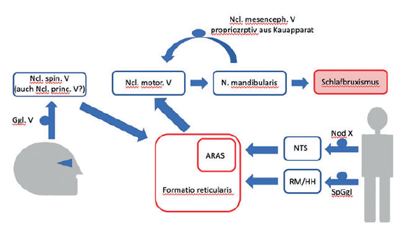
Fig. 1: Neuronal influences in sleep bruxism (from [52] with kind permission). Nod=ganglion nodosum (inferius according to new nomenclature) of the vagus. Strictly speaking, the Ggl. jugulare (superius) should also be named, since afferents from the upper aerodigestive tract, whose cell bodies are located in the Ggl. jugulare, could also trigger a microarousal. (ARAS = ascending reticular activating system).
NTS= Ncl. tractus solitarii
RM/HH= spinal cord/ posterior horn
SpGgl= Spingal ganglion
Sleep disorders – Known sleep disorders associated with bruxism include nightmares in children [4], disturbed sleep patterns in shift workers [3], insomnia [22], snoring [66] and sleep apnea [39]. It is also known that unconscious tooth clenching is most pronounced in the minutes before rapid eye movement (REM) sleep, which indicates an influence of sleep stages on SB. During sleep, physiological rhythmic masseter muscle activity (RMMA) occurs in approximately 60% of the healthy population. RMMA are defined as slow (1 Hz) chewing movements without the occurrence of tooth grinding. These rhythmic masticatory muscle activities occur about 3× more frequently and up to 1/3 more intensively in affected individuals [48]. RMMA during sleep is associated with transient activation of cortical, limbic and autonomic circuits [15]. The movements of the jaw are induced by cortical autonomic cardiac activity through the sympathetic nervous system [75]. In addition, the lobus temporale and the limbic system are activated during jaw tension with subsequent jaw clenching [15].
Psychological and behavioral risk factors – Emotional stress is discussed as an etiological (risk) factor, especially in children [34], [38]. An evidence-based statement is made difficult by the methodology of the parent survey.
The following stressors correlate with bruxism in children:
- dysfunctional family constellations [64],
- Anxiety and anxiety disorders [92],
- separated or divorced parents [12],
- working mothers [97],
- Sleep disorders [21],
- Light and/or noise in the nursery [98],
- One or both parents affected by bruxism [63] (51% of affected parents vs. 31% of parents with no history of bruxism).
In infants who are not yet able to speak, sleep bruxism can also be an expression of fear or anger [15].
SB has been associated with various characteristic personality traits:
- Anxiety and anxiety disorders [7], [23], [26], [31], [35], [36], [82], [85], [89], [92],
- Stress [31],
- Neuroticism [26],
- restless behavior (Fonseca et al. 2011)
- Fonseca CME, Dos Santos MBF, Consani RLX, et al. Incidence of sleep bruxism among children in Itanhandu, Brazil. Sleep Breath. 2011;15(2):215-220
- PTSD [15],
- Alcohol, nicotine and drug consumption; passive smoking [81],
- Excessive coffee consumption (>6 cups per day) [81],
- frequent use of dental applications [101],
- depression [70].
Socioeconomic status – Observational studies show that over half of children affected by SB have a low socioeconomic background [99].
Socioeconomic status – Observational studies show that over half of children affected by SB have a low socioeconomic background [99].
Anatomical-structural factors – Sleep bruxism can be triggered by narrowing of the airways. This can lead to sudden microarousals, which can then stimulate tachycardia. This in turn promotes rhythmic activity of the masticatory muscles and teeth grinding, which stimulates the T-cell receptors, ultimately leading to bradycardia [24].
Bruxism is also generally associated with a forward-backward displacement of the head [101].
In a recent study, Oh et al. found that the probability of occurrence of SB is increased by the risk factors enlarged tonsils, restricted tongue mobility and restricted nasal breathing [80]. Moreover, the more of the 3 risk factors are present, the higher the probability of occurrence, at 91% (3 risk factors) vs. 9% (0 risk factors).
Genotypic risk factors – Sequence variations in the genes, so-called polymorphisms, especially in the HTR2A gene, correlate with an increased serotonin-mediated risk of sleep bruxism [2], [85], [86], [103], stress and anxiety [27], [32], [42]. A systematic review also identified a genetic effect in WB. In particular, WB occurs more frequently in movement disorders associated with stereotypies [29], which are influenced by anxiety. In addition to genetic factors, epigenetic influencing factors are discussed, as syndromes such as Rett syndrome, Prader-Willi syndrome and Angelman syndrome correlate with bruxism [17].
Changes in brain biochemistry – bruxism leads to an increase in noradrenaline and catecholamine due to emotional stress [25], [96]. Furthermore, the levels of glutamate, dopamine and gamma-aminobutyric acid (GABA) increase, while these adaptations have partly positive effects [49]. Bracha et al. found that bruxism causes an increased signaling effect in the noradrenaline neurons of the truncus cerebri, especially in combination with receptors that have not yet been downregulated. Serotonin receptor subtypes play a complex role in this [15]. In particular, serotonin (5-HT) as a neurotransmitter in behavioral responses to environmental stimuli (social behavior, emotion, sleep, circadian rhythm, thermoregulation and stress coping strategies) interacts with the maintenance of arousal, regulation of stress response, muscle tone and respiration [42], [103]. This suggests that serotonin may play a role in SB pathogenesis [86]. This hypothesis is corroborated by the fact that serotonin reuptake inhibitors (SSRIs) can trigger SB episodes in susceptible individuals [61].
Reflux – Acid exposure during reflux in the esophagus initiates microarousals and rhythmic masticatory muscle activity resulting from stimulation of autonomic cortical reflexes [57]. With a reported prevalence of 74%, reflux is a common comorbidity of sleep bruxism [45].
Craniomandibular dysfunctions – As mentioned above, there is a connection between CMD and bruxism. CMD encompasses various disorders of the temporomandibular joint and masticatory system, which are characterized by pain in the temporomandibular joint or masticatory muscles, functional limitations of the jaw and associated tissues (ligaments/connective tissue structures) and clicking noises that occur during chewing (de Leeuw et al. 1994). The complexity of disorder patterns is caused by a multifactorial pathogenesis that includes traumatological, anatomical, neuromuscular and psychosocial factors (Steenks 1991, Winkelmann et al. 1999).
de Leeuw JRJ, Ros WJG, Steenls MH, et al. Multidimensional evaluation of craniomandibular dysfunction. II: Pain assessment. J Oral Rehabil. 1994;21(5):515-532
Steenks MH. Temporomandibular joint dysfunctions from a physiotherapeutic and dental point of view: diagnosis and therapy. Quintessenz-Verlag-GmbH; 1991
Winkelmann C, Schreiber TU, Weih C, et al. Approaches to physiotherapy for craniomandibular dysfunctions. Physiotherapy 1999;51:2042-2054
CMD often leads to secondary, mostly temporal headaches. Canto et al. investigated the further connection between CMD and SB. The results show a more than 3-fold increased risk of primary headache for adults with SB [18]. There is insufficient data for children to draw any conclusions. It is being discussed whether trigger points in the neck, shoulder and masticatory muscles lead to referred tension headache by triggering central sensitization processes. One hypothesis is that headaches are induced via nociceptors in the masticatory muscles and temporomandibular joints. If bruxism then occurs, this could lead to a reduction in the stimulation threshold of the nucleus spinalis nervi trigemini pars caudalis or to a central sensitization of this nerve area [18].
Pathophysiology of sleep bruxism
Peripheral occlusal factors (e.g. tooth occlusion as a morphological factor) play a subordinate role in contrast to central regulation [59], [104] (see above). In central regulation, a distinction is also made between pathophysiological and psychosocial factors [104]. Pathophysiological factors include abnormal excitability of the central motor innervation of the jaw. This may be due to incorrectly regulated brainstem inhibitory circuits, e.g. from cranial nerves V and X, as well as subcortical structures [33]. The psychosocial risk factors have been described above.
During sleep, pathophysiological variations of nerve patterns occur through the (para)sympathetic nervous system [79]. These different excitation patterns are shown in Fig. 2, the temporal sequence is summarized in Fig. 3.
Fig. 2: Influence of the sympathetic (SNS) and parasympathetic (PSNS) nervous system on the course of sleep bruxism (according to [79]):
- Pattern A: Activation of the SNS induces SB activity, which is continued by PSNS activation. The mean onset time of the SNS (before SB activity) is 11.8 sec; the mean onset time of the PSNS (after onset of SB activity) is 10.3 sec.
- Pattern B: Activation of the SNS induces SB activity, which is not followed by activation of the PSNS.
- Pattern C: The self-service activation takes place without the SNS, but is accompanied by the PSNS.
- Pattern D: The self-service activity starts without the involvement of the SNS or PSNS.
Fig. 3: Representation of the temporal relationship between the mean onset time of SNS and PSNS and the occurrence of sleep bruxism in seconds (according to [79])
Neuroevolutionary and paleoanthropological perspectives
In the course of evolution (approx. 2 million years to 200,000 years ago), bruxism led to a strengthening of the masseter and temporal muscles and thus also of the bite. Especially in the time before the discovery of fire, this was fundamental for the feeding of plants and led to the survival of Homo [15].
In 2005, Bracha also hypothesized that jaw clenching leads to increased blood flow through the partes mastiodea of the temporal bone, which may lead to increased blood flow through the temporal lobe structures involving acute activation of limbic fear circuits [15]. Thus, an alternative stress-induced fear circuit, so-called freezing or slackening by vagus activation, may have evolved as a possible survival advantage as opposed to the flight-or-fight response [13], [16]. Of course, a strong bite also served as a defense, and human oral flora combined with a strong bite produced a high mortality rate in prey and attack [90]. These aspects of bruxism, which can certainly be seen as positive, continued in the adaptive purpose of keeping the teeth sharp [15].
Thus, bruxism in modern times can also be seen as a manifestation of acute anxiety or chronic emotional stress [15].
Further advantages of bruxism
On the one hand, SB could represent a protective function at night [41], [58], e.g. in the maintenance of respiration by supporting the masseter for the genioglossus muscle. This acts as an efficient dilator of the upper airway [46], which leads to lubrication of the oropharynx [76] and thus protects the upper section of the digestive tract [49], [76], [100]. On the other hand, maximum masseter activity leads to improved performance in golf [94]. In addition, a strong bite serves as self-defense in the event of physical and sexual assault [90].
Potential consequences of bruxism
In children, SB rarely requires intervention [74] and usually does not result in serious damage [91]. The most important consequences that can nevertheless occur include tooth destruction, tooth loss, temporomandibular joint and jaw muscle pain, restriction of jaw movement and primary headaches (in 60% of SB sufferers) [8], [40], [44], [93]. In healthy children, the figure is only 31% [12]. However, SB can lead to stress-induced disorders such as chronic fatigue syndrome and fibromyalgia [1]. Furthermore, there are correlations, but no causalities, between gastroesophageal reflux disease, depression, anxiety disorders and reduced sleep quality with bruxism [50].
Diagnosis of sleep bruxism
The following symptoms and signs are described by patients [77]:
- Pain in the jaw joints, chewing and neck muscles,
- Headaches, especially in the temple area on waking,
- Hypersensitive teeth,
- Excessive tooth mobility without an identifiable dental reason,
- poor sleep.
The following clinical signs can be found [77]:
- non-carious loss of tooth structure and/or
- Loss of restorative materials, reconstructions or fillings,
- Tongue and cheek impressions,
- whitish keratinization ridge in the buccal planum,
- localized gum recession,
- Hypertonus of the masticatory muscles,
- Jaw opening and closing restricted,
- Torus palatinus or tori mandibulares,
- Pressure sensitivity and a visible enlargement of the temporalis and masseter muscles [15].
Furthermore, bruxism can be categorized more or less reliably using various diagnostic approaches (mod. after [88]).
- possible WB/SB: positive indications from survey/questionnaires,
- probable WB/SB: positive clinical evidence with or without positive evidence from interview/questionnaires,
- definitive WB/SB: positive instrumental findings with or without positive findings from interviews/questionnaires and/or positive clinical signs.
An (early) diagnosis to reduce clinical signs is of great importance. Non-carious tooth structure loss, loss of restorative materials, periodontal problems, the development of CMD and headaches should be avoided [88].
The American Association of Sleep Medicine (AASM) has already established criteria for the diagnosis of SB (Table 1) [6].

Table 1: American Association for Sleep Medicine (AASM) criteria for sleep-associated bruxism
Treatment approaches
As highlighted above, sleep bruxism serves adaptive responses and protective mechanisms. Several critical systematic reviews found very weak or no evidence for the association between SB and negative health outcomes [59], [72], [73]. Thus, Beddis concludes that it is unlikely that bruxism has no function, and therefore treatment should only take place in cases of problems due to SB [10]. As SB rarely leads to negative effects in children, manual therapy treatment strategies in this age group are controversial [95].
Manual treatment approaches
Reduction of muscle hypertonus – by massaging and/or stretching the masticatory and neck muscles, e.g. using muscle energy techniques, can increase the range of movement, reduce pain and improve the resting position of the lower jaw [102].
Abdominal breathing – In general, diaphragmatic breathing can be trained right into the abdomen; if necessary, diaphragmatic dysfunctions and associated organ dysfunctions (e.g. stomach, liver, lungs, heart, etc.) can be corrected.
Promotion of relaxation – through relaxing activities, e.g. by visualizing images or consciously relaxing various muscle groups [84] or through vagus-stimulating interventions [55], [56].
LEIT reset – Extrinsic and intrinsic jaw techniques are applied in a coordinated manner (Liem extrinsic and intrinsic TMG reset):
Extrinsic techniques performed by practitioners:
- Myofascial release techniques and trigger point treatment: especially of the masticatory, hyoid, neck and shoulder muscles.
- Techniques for the temporomandibular joint and associated structures such as the ligament-capsule apparatus [52].
- Posture: treatment of ascending and descending muscle chains [52].
- Neuromuscular inhibition techniques: e.g. of the ascending reticular activating system (ARAS), afferents of the cranial nerves (especially V, VII, IX, X), the posterior horn in the spinal cord and somatic nerves.
- Techniques for the autonomic nervous system: e.g. inhibition techniques of the sympathetic nervous system and vagus nerve stimulation techniques [55], [56].
- For reflux: technique for the lower esophageal sphincter [28].
- If the airways are narrowed: mobilization of the thorax and airways and breathing techniques [9].
- For enlarged tonsils: lymphatic stimulation of the throat, head and neck region [51], [52], dietary changes [56].
- If tongue mobility is restricted: treatment of dyskinesia, tongue mobilization [52].
- For emotional stress, anxiety: treatment using psychosomatic osteopathy approaches, e.g. multimodal bifocal integration [53], [54].
Intrinsic self-help techniques carried out by those affected:
- Chewing, hyoid and neck muscle techniques, e.g. with alternating tension and relaxation or pure muscle relaxation exercises such as “jaw drop” and/or reciprocal muscle tension inhibition techniques [52].
- Breathing exercises: especially slowed abdominal breathing with the tongue on the palate [56].
- Posture exercises, e.g. to correct the head/neck position.
- Myofascial vibration techniques while lying and sitting.
- Restricted tongue mobility and enlarged tonsils: Use of the FaceFormer: Patients who had trained with the FaceFormer were compared with those who had been fitted with grinding splints. There was no improvement in splint patients, but rather an increase in grinding/clenching, while the FaceFormer group showed a significant improvement after just 8 weeks [11].
- Relaxation exercises, vagus nerve-stimulating self-exercises and HRV (heart rate variability) biofeedback [56].
- Anxiety-reducing self-exercises.
Literature on:
Liem T. Bruxism and osteopathy. Osteopathic Medicine 2022; 2nd ed:
Literature
- Aaron LA, Buchwald D. A review of the evidence for overlap among unexplained clinical conditions. Ann Intern Med. 2001; 134 (9_Part_2): 868-881
- Abe Y, Suganuma T, Ishii M et al. Association of genetic, psychological and behavioral factors with sleep bruxism in a Japanese population. J Sleep Res. 2012; 21 (3): 289-296
- Ahlberg K, Jahkola A, Savolainen A et al. Associations of reported bruxism with insomnia and insufficient sleep symptoms among media personnel with or without irregular shift work. Head Face Med. 2008; 4 (1): 1-6
- Alencar NA de, Fernandes ABN, Souza MMG de et al. Lifestyle and oral facial disorders associated with sleep bruxism in children. CRANIO®. 2017; 35 (3): 168-174
- Alfano CA, Bower JL, Meers JM. Polysomnography-detected bruxism in children is associated with somatic complaints but not anxiety. J Clin Sleep Med. 2018; 14 (1): 23-29
- American Association of Sleep Medicine (AASM). International classification of sleep disorders. Diagnostic coding Man. Published online 2005: 51-55
- Alves AC, Alchieri JC, Barbosa GAS. ACTA-1-2013-final_3-2011. published online 2013
- Bader G, Lavigne G. Sleep bruxism; an overview of an oromandibular sleep movement disorder. Sleep Med Rev. 2000; 4 (1): 27-43
- Benjamin JG, Moran RW, Plews DJ, Kilding AE, Barnett LE, Verhoeff WJ, Bacon CJ. The effect of osteopathic manual therapy with breathing retraining on cardiac autonomic measures and breathing symptoms scores: A randomized wait-list controlled trial. J Bodyw Mov Ther. 2020 Jul; 24 (3): 282-292
- Beddis H, Pemberton M, Davies S. Sleep bruxism: an overview for clinicians. Br Dent J. 2018; 225 (6): 497-501
- Berndsen KJ, Berndsen S, Kopp ST. New ways of functional treatment of bruxism Face Former Therapy. Journal Orofacial Orthopedics. 2009; 70: 420
- Bortoletto CC, Salgueiro M da CC, Valio R et al. The relationship between bruxism, sleep quality, and headaches in schoolchildren. J Phys Ther Sci. 2017; 29 (11): 1889-1892
- Bracha BHS. 2004_bracha -FEAR–Freeze_Flight_Fight_Fright_Faint. 2004; 9 (9): 679-685
- Bracha HS, Bracha AS, Williams AE et al. The human fear-circuitry and fear-induced fainting in healthy individuals: The paleolithic-threat hypothesis. Clin Auton Res. 2005; 15 (3): 238-241
- Bracha HS, Ralston TC, Williams AE et al. The clenching-grinding spectrum and fear circuitry disorders: Clinical insights from the neuroscience/paleoanthropology interface. CNS Spectr. 2005; 10 (4): 311-318
- Bracha HS, Ralston TC, Matsukawa JM et al. Does “Fight or Flight” Need Updating? 2004; (October)
- Čalić A, Peterlin B. Epigenetics and Bruxism: Possible Role of Epigenetics in the Etiology of Bruxism. Int J Prosthodont. 2015; 28 (6)
- Canto G, Singh V, Bigal M et al. Association Between Tension-Type Headache and Migraine with Sleep Bruxism: A Systematic Review. Headache. 2014; 54
- Carlsson GE, Egermark I, Magnusson T. Predictors of bruxism, other oral parafunctions, and tooth wear over a 20-year follow-up period. J Orofac Pain. 2003; 17 (1)
- Castrillon EE, Ou K-L, Wang K et al. Sleep bruxism: an updated review of an old problem. Acta Odontol Scand. 2016; 74 (5): 328-334
- Castroflorio T, Bargellini A, Rossini G et al. Risk factors related to sleep bruxism in children: A systematic literature review. Arch Oral Biol. 2015; 60 (11): 1618-1624
- Castroflorio T, Bargellini A, Rossini G et al. Sleep bruxism in adolescents: a systematic literature review of related risk factors. Eur J Orthod. 2017; 39 (1): 61-68
- Cheifetz AT, Osganian SK, Allred EN et al. Associated Correlates in Children as Reported by Parents. J Dent Child. 2005; 72 (2): 67-73
- Chowdhury T, Bindu B, Singh GP et al. Sleep disorders: Is the trigemino-cardiac reflex a missing link? Front Neurol. 2017; 8 (FEB): 1-7
- Clark GT, Rugh JD, Handelman SL. Nocturnal Masseter Muscle Activity and Urinary Catecholamine Levels in Bruxers. J Dent Res. 1980; 59 (10): 1571-1576
- Cruz-Fierro N, Martínez-Fierro M, Cerda-Flores RM et al. The phenotype, psychotype and genotype of bruxism. Biomed Reports. 2018; 8 (3): 264-268
- Donnellan MB, Burt SA, Levendosky AA et al. Genes, personality, and attachment in adults: A multivariate behavioral genetic analysis. Personal Soc Psychol Bull. 2008; 34 (1): 3-16
- Eguaras N, Rodríguez-López ES, Lopez-Dicastillo O, Franco-Sierra MÁ, Ricard F, Oliva-Pascual-Vaca Á. Effects of Osteopathic Visceral Treatment in Patients with Gastroesophageal Reflux: A Randomized Controlled Trial. J Clin Med. 2019 Oct 19; 8 (10): 1738
- Ella B, Ghorayeb I, Burbaud P et al. Bruxism in movement disorders: a comprehensive review. J Prosthodont. 2017; 26 (7): 599-605
- Every RG. Significance of tooth sharpness for mammalian, especially primate, evolution. Contrib Primatol. 1975; 5: 293-325
- Ferreira-Bacci A do V, Cardoso CLC, Díaz-Serrano KV. Behavioral problems and emotional stress in children with bruxism. Braz Dent J. 2012; 23 (3): 246-251
- Fraley RC, Roisman GI, Booth-LaForce C et al. Interpersonal and genetic origins of adult attachment styles: a longitudinal study from infancy to early adulthood. J Pers Soc Psychol. 2013; 104 (5): 817
- Gastaldo E, Member S, Quatrale R et al. Bruxism Centrally Mediated-not occlusion. Published online 2005: 145-155
- Giraki M, Schneider C, Schäfer R et al. Correlation between stress, stress-coping and current sleep bruxism. Head Face Med. 2010; 6 (1)
- Gorayeb MAM, Gorayeb R. Cefaléia associada a indicadores de transtornos de ansiedade em uma amostra de escolares de Ribeirão Preto, SP. Arq Neuropsiquiatr. 2002; 60 (3B): 764-768
- Gungormus Z, Erciyas K. Evaluation of the relationship between anxiety and depression and bruxism. J Int Med Res. 2009; 37 (2): 547-550
- Guo H, Wang T, Niu X et al. The risk factors related to bruxism in children: A systematic review and meta-analysis. Arch Oral Biol. 2018; 86: 18-34
- Insana SP, Gozal D, McNeil DW et al. Community based study of sleep bruxism during early childhood. Sleep Med. 2013; 14 (2): 183-188
- Jokubauskas L, Baltrušaitytė A. Relationship between obstructive sleep apnea syndrome and sleep bruxism: a systematic review. J Oral Rehabil. 2017; 44 (2): 144-153
- Kato T, Lavigne GJ. Sleep Bruxism: A Sleep-Related Movement Disorder. Sleep Med Clin. 2010; 5 (1): 9-35
- Khoury S, Rouleau GA, Rompré PH et al. A significant increase in breathing amplitude precedes sleep bruxism. Chest. 2008; 134 (2): 332-337
- Kiser D, SteemerS B, Branchi I et al. The reciprocal interaction between serotonin and social behavior. Neurosci Biobehav Rev. 2012; 36 (2): 786-798
- Kleinberg I. Bruxism: aetiology, clinical signs and symptoms. Aust Prosthodont J. 1994; 8: 9-17
- Knutson GA. Vectored upper cervical manipulation for chronic sleep bruxism, headache, and cervical spine pain in a child. J Manipulative Physiol Ther. 2003; 26 (6): 395
- Kuhn M, Türp JC Risk factors for bruxism-A literature review from 2007 to 2016 Swiss Dent J. 2018; 128: 125-129
- Landry M-L, Rompré PH, Manzini C et al. Reduction of sleep bruxism using a mandibular advancement device: an experimental controlled study. Int J Prosthodont. 2006; 19 (6)
- Lavigne GJ, Khoury S, Abe S et al. Bruxism physiology and pathology: An overview for clinicians. J Oral Rehabil. 2008; 35 (7): 476-494
- Lavigne GJ, Rompré PH, Poirier G et al. Rhythmic masticatory muscle activity during sleep in humans. J Dent Res. 2001; 80 (2): 443-448
- Lavigne GJ, Introduction I. Neurobiological mechanisms involved in sleep bruxism. Crit Rev Oral Biol Med. 2003; 14 (1): 30-46
- Li Y, Yu F, Niu L et al. Association between bruxism and symptomatic gastroesophageal reflux disease: A case-control study. J Dent. 2018; 77 (July): 51-58
- Liem T. Update on drainage of the brain and osteopathic treatment approaches, Osteopathische Medizin 2017; 18 (2), 22-27
- Liem T. The practice of craniosacral osteopathy. 4th revised and expanded edition. Thieme, Stuttgart, 2020
- Liem T. Neuhuber W. Osteopathic Treatment Approach to Psychoemotional Trauma by Means of Bifocal Integration: clinical practice, Journal of the American Osteopathic Association 2020a; 120 (3), 1-10
- Liem T. Neuhuber W. Psychosomatic osteopathy in trauma using the example of bifocal integration. Osteop Med. 2020b; 21 (4): 6-13
- Liem T. Vagus activation and stress response from an osteopathic perspective. Osteopathic Medicine. 2021; 22 (4): 10-15
- Liem T, Tsolodimos C. The Osteopathy Self-Help Book. How osteopathy works and promotes self-healing. With exercises for every day. Trias, Stuttgart; 2022
- Lobbezoo F, Ahlberg J, Manfredini D et al. Are bruxism and the bite causally related? J Oral Rehabil. 2012; 39 (7): 489-501
- Lobbezoo F, Ahlberg J, Glaros AG et al. Bruxism defined and graded: An international consensus. J Oral Rehabil. 2013; 40 (1): 2-4
- Lobbezoo F, Ahlberg J, Raphael KG et al. International consensus on the assessment of bruxism: Report of a work in progress. J Oral Rehabil. 2018; 45 (11): 837-844
- Lobbezoo F, Rompré PH, Soucy JP et al. Lack of Associations between Occlusal and Cephalometric Measures, Side Imbalance in Striatal D2 Receptor Binding, and Sleep-Related Oromotor Activities. J Orofac Pain. 2001; 15 (1): 64-71
- Lobbezoo F, Van Denderen RJA, Verheij JGC et al. Reports of SSRI-Associated Bruxism in the Family Physician’s Office. J Orofac Pain. 2001; 15 (4): 340-346
- Lobbezoo F, van der Zaag J, Naeije M. Bruxism: its multiple causes and its effects on dental implants-an updated review. J Oral Rehabil. 2006; 33 (4): 293-300
- Lobezzo F, Visscher CM, Ahlberg J et al. Bruxism and genetics: a review of the literature -. J Oral Rehabil. 2014; 41: 709-714
- Lurie O, Zadik Y, Einy S et al. Bruxism in military pilots and non-pilots: tooth wear and psychological stress. Aviat Space Environ Med. 2007; 78 (2): 137-139
- Machado E, Dal-Fabbro C, Cunali PA et al. Prevalence of sleep bruxism in children: A systematic review. Dental Press J Orthod. 2014; 19 (6): 54-61
- Maluly M, Andersen ML, Dal-Fabbro C et al. Polysomnographic study of the prevalence of sleep bruxism in a population sample. J Dent Res. 2013; 92 (7_suppl): S97-S103
- Manfredini D, Arreghini A, Lombardo L et al. Assessment of anxiety and coping features in bruxers: a portable electromyographic and electrocardiographic study. J Oral Facial Pain Headache. 2016; 30 (3): 249-254
- Manfredini D, Bracci A, Djukic G. BruxApp: the ecological momentary assessment of awake bruxism. Minerva Stomatol. 2016; 65 (4): 252-255
- Manfredini D, Bucci MB, Sabattini VB et al. Bruxism: overview of current knowledge and suggestions for dental implants planning. CRANIO®. 2011; 29 (4): 304-312
- Manfredini D, Fabbri A, Peretta R et al. Influence of psychological symptoms on home-recorded sleep-time masticatory muscle activity in healthy subjects. J Oral Rehabil. 2011; 38 (12): 902-911
- Manfredini D, Lobbezoo F. Role of psychosocial factors in the etiology of bruxism. J Orofac pain. 2009; 23 (2): 153-166
- Manfredini D, Lobbezoo F. Relationship between bruxism and temporomandibular disorders: A systematic review of literature from 1998 to 2008. Oral Surgery, Oral Med Oral Pathol Oral Radiol Endodontology. 2010; 109 (6): e26-e50
- Manfredini D, Poggio CE, Lobbezoo F Is bruxism a risk factor for dental implants? A systematic review of the literature. Clin Implant Dent Relat Res. 2014; 16 (3): 460-469
- Manfredini D, Restrepo C, Diaz-Serrano K et al. Prevalence of sleep bruxism in children: a systematic review of the literature. J Oral Rehabil. 2013; 40 (8): 631-642
- Marthol H, Reich S, Jacke J et al. Enhanced sympathetic cardiac modulation in bruxism patients. Clin Auton Res. 2006; 16 (4): 276-280
- Miyawaki S, Lavigne GJ, Mayer P et al. Association between sleep bruxism, swallowing-related laryngeal movement, and sleep positions. Sleep. 2003; 26 (4): 461-465
- Murali R V, Rangarajan P, Mounissamy A Bruxism: Conceptual discussion and review. J Pharm Bioallied Sci. 2015; 7 (Suppl 1): S265
- Murray CG, Sanson GD. Thegosis-A critical review. Aust Dent J. 1998; 43 (3): 192-198
- Nukazawa S, Yoshimi H, Sato S. Autonomic nervous activities associated with bruxism events during sleep. CRANIO®. 2018; 36 (2): 106-112
- Oh JS, Zaghi S, Ghodousi N et al. Determinants of probable sleep bruxism in a pediatric mixed dentition population: a multivariate analysis of mouth vs. nasal breathing, tongue mobility, and tonsil size. Sleep Med. 2021; 77: 7-13
- Ohayon MM, Li KK, Guilleminault C. Risk factors for sleep bruxism in the general population. Chest. 2001; 119 (1): 53-61
- de Oliveira MT, Bittencourt ST, Marcon K et al. Sleep bruxism and anxiety level in children. Braz Oral Res. 2015; 29 (1): 1-5
- Olmos SR. Comorbidities of chronic facial pain and obstructive sleep apnea. Curr Opin Pulm Med. 2016; 22 (6): 570-575
- Ommerborn MA, Schneider C, Giraki M et al. Effects of an occlusal splint compared with cognitive-behavioral treatment on sleep bruxism activity. Eur J Oral Sci. 2007; 115 (1): 7-14
- Oporto Gh, Bornhardt T, Iturriaga V et al. Genetic polymorphisms in the serotonergic system are associated with circadian manifestations of bruxism. J Oral Rehabil. 2016; 43 (11): 805-812
- Oporto GH, Lagos JD, Bornhardt T et al. ¿ Es posible la contribución de factores genéticos en el bruxismo? Int J Odontostomatol. 2012; 6 (3): 249-254
- Ortiz MI, Rangel-Barragán RO, Contreras-Ayala M et al. Procedural pain and anxiety in pediatric patients in a Mexican dental clinic. Oral Health Dent Manag. 2014; 13 (2): 495-501
- Peroz I, Bernhardt O, Kares H et al. S3 guideline (long version) Diagnosis and treatment of bruxism. Journal of Craniomandibular Function. 2019; 11 (3): 225-i385
- Petit D, Touchette É, Tremblay RE et al. Dyssomnias and parasomnias in early childhood. Pediatrics. 2007; 119 (5)
- Pretty IA, Hall RC. Forensic dentistry and human bite marks: issues for doctors. Hosp Med. 2002; 63 (8): 476-482
- Reddy SV, Kumar M, Sravanthi D. Bruxism: A Literature Review. J Int Oral Heal. 2014; 6 (6): 105-109
- Restrepo CC, Vásquez LM, Alvarez M et al. Personality traits and temporomandibular disorders in a group of children with bruxing behavior. J Oral Rehabil. 2008; 35 (8): 585-593
- Restrepo C, Gómez S, Manrique R. Treatment of bruxism in children: a systematic review. Quintessence Int. 2009; 40 (10): 849-855
- Ringhof S, Hellmann D, Meier F et al. The effect of oral motor activity on the athletic performance of professional golfers. Front Psychol. 2015; 6 (JUN): 1-10
- Santos C, Amorim M, Ferreira E et al. Ejjectivenesss of two. Published online 2014: 1-8
- Seraidarian P, Seraidarian PI, das Neves Cavalcanti B et al. Urinary levels of catecholamines among individuals with and without sleep bruxism. Sleep Breath. 2009; 13 (1): 85-88
- Seraj B, Shahrabi M, Ghadimi S et al. The prevalence of bruxism and correlated factors in children referred to dental schools of Tehran, based on parent’s report. Iran J Pediatr. 2010; 20 (2): 174
- Serra-Negra JM, Paiva SM, Fulgêncio LB et al. Environmental factors, sleep duration, and sleep bruxism in Brazilian schoolchildren: a case-control study. Sleep Med. 2014; 15 (2): 236-239
- Serra-Negra JM, Paiva SM, Seabra AP et al. Prevalence of sleep bruxism in a group of Brazilian schoolchildren. Eur Arch Paediatr Dent. 2010; 11 (4): 192-195
- Thie NMR, Kato T, Bader G et al. The significance of saliva during sleep and the relevance of oromotor movements. Sleep Med Rev. 2002; 6 (3): 213-227
- Vélez AL, Restrepo CC, Peláez-Vargas A et al. Head posture and dental wear evaluation of bruxist children with primary teeth. J Oral Rehabil. 2007; 34 (9): 663-670
- Visscher CM, Lobbezoo F, Naeije M. Treatment of bruxism: physiotherapeutic approach. Ned Tijdschr Tandheelkd. 2000; 107 (7): 293-296
- Wilhelm K, Siegel JE, Finch AW et al. The long and the short of it: Associations between 5-HTT genotypes and coping with stress. Psychosom Med. 2007; 69 (7): 614-620
- Yap AUJ, Chua AP. Sleep bruxism: Current knowledge and contemporary management. J Conserv Dent. 2016; 19 (5): 383-389