A recent study shows that the factors influencing the development and morphology of the tentorium cerebelli differ particularly between the early and mid-fetal periods.
In the early fetal period, the cerebrum covers about half of the midbrain. The separation of the dural limiting layer in the parieto-occipital region extends from the posterior cerebrum to the cranial cerebellum. The lateral folds of the tentorium cerebelli were distributed between their apex, which coincides with the falx cerebri, and their basal plane, located between the midbrain and the rostral hindbrain. The differences in the growth directions of the tentorium cerebelli components gradually decrease as the cerebrum covers the midbrain. Rotation of the tentorium cerebelli in its middle portion was demonstrated, consistent with its growth, which ceased in the mid-fetal period. The brainstem and cerebellum extend downward through differential growth, while the cerebrum covers them superiorly. The morphology of the tentorium cerebelli curved to adapt to the surfaces of the cerebellum and cerebrum. A study by Matsunari et al. (2022) suggests that the morphology of the tentorium cerebelli is influenced by factors that differ particularly between the early and mid-fetal periods. The present data from Matsunari et al. (2022) provide a much more comprehensive picture of tentorium cerebelli development depending on the fetal developmental stage than has previously been presented. 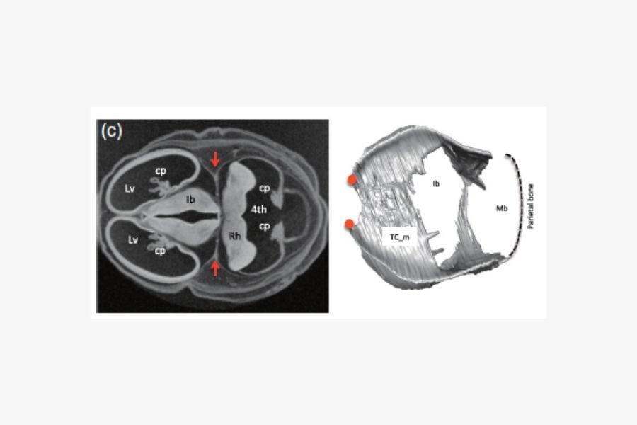

References:
Matsunari C, Kanahashi T, Otani H, Imai H, Yamada S, Okada T, Takakuwa T. Tentorium cerebelli formation during human embryonic and early fetal development. Anat Rec (Hoboken). 2023 Mar;306(3):515-526.