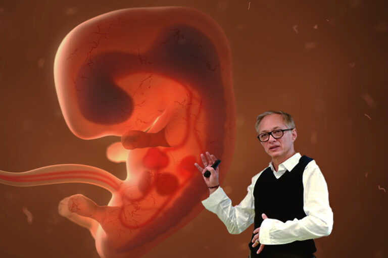
Leonardo Vieira, a Brazilian professor of osteopathic medicine, recently published a review on the human fascial system, in which he presents the relationship between the embryonic origin of the musculoskeletal, visceral, pannicular, and neural fasciae and the neuronal system. A working group of the Fascia Research Society defines the term “fascial system” as follows: “The fascial system consists of a three-dimensional continuum of soft connective tissue containing fibrous, loose, and dense collagen that permeates the body. It includes elements such as adipose tissue, adventitia, neurovascular sheaths, aponeuroses, deep and superficial fasciae, epineurium, joint capsules, ligaments, membranes, meninges, myofascial expansions, periosteum, retinacula, septa, tendons, visceral fasciae, and intermuscular connective tissue, including endomysium/epimysium/perimysium. The fascial system incorporates all organs, muscles, bones, and nerve fibers, interweaving and permeating them, thereby providing the body with a functional structure and creating an environment that enables all systems in the body to work in an integrated manner” (Schleip, Hedley, Yucesoy, 2019).
Somitogenesis
Somite formation begins on the twentieth day and continues until all 44 pairs have been formed. The somites are formed from the paraxial mesoderm and can be divided into the following structures:
- Sclerotome
- Bones of the body (except cranial bones and hyoid bone)
- Dermatome
- Dermis, superficial fasciae, layers of subcutaneous adipose tissue
- Innervation by anterior and posterior rami of the spinal nerve
- Myotome
- Muscles, deep fascia (except face and anterior neck)
- Subdivision into:
- Epimere: dorsal fasciae and muscles of the trunk (erector spinae muscles)
- Hypomere: ventral muscles and fasciae of the trunk, limbs, deep fascia with its three layers (except Mm. trapezius and sternocleidomastoideus)
The human fascial system is consequently formed primarily from the paraxial mesoderm. The exception to this is the development of the facial fasciae and those of the ventral cervical region, which develop from mesenchymal cells derived from the ectodermally-derived neural crest. In this process, the differentiation of the neural tube and the development of the fascial system mutually influence each other, creating a close connection. This is referred to as the neurofascial system. However, the author explicitly emphasizes that the fascial system cannot be linearly divided into different models, but rather, at the microscopic level, surrounds the entire body as a large fascial network. Thus, it represents an efficient communication system for the body (Vieira, 2020). References: Schleip R, Hedley G, Yucesoy CA. Fascial nomenclature: Update on related consensus process. Clin Anat. 2019;32(7):929-933. Vieira L. Embryology of the Fascial System. Cureus. 2020;12(8):1-21.