Contents
From: Liem T. Craniosacral Osteopathy, 2018; 7th rev. ed. Thieme, Stuttgart, 420ff.
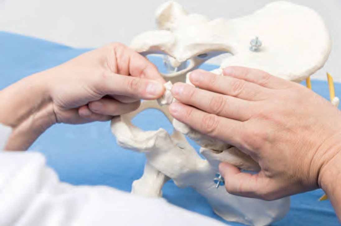
Patient
- in side-lying position, hips and knees slightly bent
Hand position
- Gently insert the index or middle finger into the patient’s anus and place the thumb of the same hand externally on the coccyx.
- The coccyx is grasped by both fingers.
- The cranial hand is placed on the sacrum or, depending on the palpable tension, encompasses other surrounding structures, e.g., lumbar vertebral bodies, the skin surface of the thoracolumbar fascia, the sacrotuberous ligament, the sacrospinous ligament, the gluteus maximus muscle, the coccygeus muscle.
Execution
- With a slight caudal traction on the coccyx, follow the bone’s inherent movements.
- Lesion patterns can be treated by targeted stretching between structures.
- In accordance with techniques for treating transverse diaphragms, only repetitive movements are prevented to free the joint connections from their abnormal tension patterns.
- Additionally or alternatively, the coccyx can be gently moved in the direction of restricted movement using a direct technique.
- Instead of traction, the structures can also be gently compressed towards each other.
- Subsequently, the tissue’s reaction to this compression field is perceived and supported.
- Local ligamentous structures can be treated directly by firm, targeted pressure with the thumb on ligamentous and osseous structures. This is often indicated for the posterior sacroiliac ligaments, but also for the sacrococcygeal ligaments.
- For deep, linearly occurring pain or dysfunction, the fascial structures can be slowly and deeply stroked with the thumb, following the course of the pain.
- Paraspinal muscles, as well as those above and below the filum terminale, are inhibited by gentle pressure and myofascial following.
These procedures can also be used for targeted treatment of the dorsal ligamentous and fascial system, as well as the structures from the coccyx to the epidural space in the lumbosacral junction.
Tension balance from the coccyx to the epidural space in the lumbosacral junction
- Dural tensions are perceived from the coccyx via the filum terminale, the lumbosacral ligament, and the anterior sacrodural ligament (of Trolard).
Note: The transmission of tension from the dura to the intervertebral discs decreases with age, as the lumbosacral ligament becomes increasingly porous.
- A BLT is established between the coccyx and the lumbar vertebra via the lumbosacral ligament.
- From the coccyx, tensions of the posterior longitudinal ligament can be perceived via the filum terminale externum through the deep posterior sacrococcygeal ligaments.
- Alternatively, tensions from the coccyx are palpated via the filum terminale externum and the superficial tendinous plate of the superficial posterior sacrococcygeal ligaments.
- In addition, the osteopath perceives dural tensions via the dorsolateral dural ligaments (of Hofmann) through the contact between the sacrum and the lumbar vertebral bodies.
- Palpation of ligamentous tensions between the coccyx and the levator ani muscle, coccygeus muscle, and external anal sphincter muscle via the superficial and deep posterior sacrococcygeal ligaments through the anococcygeal attachment.
- Caudal traction is performed only to the extent that the coccyx is moved away from its tension area. Tissue barriers are not confronted.