Summary
Studies on the existence of regular lymphatic vessels in the dural sinuses, the blood-brain barrier and the immune privilege of the CNS lead to new clinical conclusions. Based on the new findings on cerebrospinal fluid research and drainage of the brain, possible osteopathic approaches are presented, e.g. heart-focused palpation, osteopathic “felt sense”, techniques for cisterns, compression of the 3rd and 4th ventricles (e.g. the ventricles of the brain). and 4th ventricles (CV-3, CV-4), compression of the lateral ventricles, dura techniques, lymphatic pump, OA release, osteopathic lymphatic drainage, drainage of the nose, eye, ear, cranial nerves and upper cervical nerves.
Keywords
Blood-brain barrier, cerebrospinal fluid, lymph vessels, dural sinus, osteopathic manipulative treatment, osteopathic lymph drainage
Keywords
Blood brain barrier, cerebrospinal fluid, lymphatic channel, dural sinus, osteopathic manipulative treatment (OMT), osteopathic lymph drainage
Blood-brain barrier with a special focus on the pericytes
According to Sá-Pereira et al. (2012), the blood-brain barrier (BBB) is a complex and dynamic interface composed of different cells that form a functional unit, the neurovascular unit (NVE). These include endothelial cells, the basement membrane, astrocytes, pericytes and neurons. It is possible that microglia and oligodendrocytes also influence the function of the BBB and thus neurodegenerative and immunological processes (Sá-Pereira et al. 2012).Pericytes are not only significantly involved in the maintenance and stabilization of the BBB, but also appear to play an important role in the development of blood vessels. Pericytes also have contractile elements that may influence blood circulation in the brain microvasculature. In addition, Sá-Pereira et al. (2012) report on possible immunological and phagocytotic influences as well as the role of pericytes in homeostasis. Several studies have shown that pericytes are functionally multipotent stem cells.
Lymph vessels in the brain
Until 2015, the state of research was that the CNS lacked a classic lymphatic drainage system. However, a study by Louveau et al. (2015) was able to show that regular lymphatic vessels exist in the meningeal membranes outside the brain parenchyma. Although there was now a consensus that the CNS is under constant immune surveillance, which takes place in the meningeal compartment, the control mechanisms for the entry and exit of immune cells in the CNS remained unclear. In 2015, while searching for entry and exit ports for T cells in the meninges, researchers from the School of Medicine at the University of Virginia discovered functional lymphatic vessels lining the dural sinuses (National Institutes of Health 2015) (Figs. 1 and 2).The vascular structures show all the molecular characteristics of lymphatic endothelial cells. These lymphatic vessels carry waste products with cerebrospinal fluid (LCS) drainage from the glymphatic system. They can transport fluid and immune cells from the cerebrospinal fluid and are connected to the deep cervical lymph nodes. However, it is also known that cardiac pulsation does not provide more than 15-25% of the driving energy, consequently other mechanisms must be responsible for the convective LCS flow dynamics (Kiviniemi et al. 2016).The complex anatomical architecture of the Virchow-Robin space (VRS, Fig. 3) allows bidirectional fluidic exchange between the VRS and the extracellular space of the brain as well as the subarachnoid space (blue arrows). The membranes of the glial cells (blue lines) and those of the pia mater (yellow lines) surround the VRS and thus control the fluid exchange. It should be noted that there is no consensus as to whether the VRS represents an open, fluid-filled space. Both experimental and clinical evidence suggest a pathway for drainage of the ISF along the basement membranes of capillaries, arterioles and arteries that leads into the lymphatic system (red lines and green arrows). The significance of the perivascular spaces surrounding the arteries and veins below the pia mater (light blue) remains unclear. They may serve as additional drainage pathways. It is also under discussion whether the glymphatic pathways connect the arterial and venous Virchow-Robin spaces with the venous perivascular spaces (black arrows) (Brinker et al. 2014).Fig. 1: Overview of the lymphatic system: old ( a) and an update (b) to illustrate the new findings. © University of Virginia Health System From: https://www.nih.gov/news-events/nih-research-matters/lymphatic-vessels-discovered-central-nervous-systemAbb. 2: Schematic representation of the intrameningeal lymphatic network newly discovered by Aleksander Aspel and colleagues. a It was known that the lymphatic vessels of the nasal mucosa were involved in the drainage of cerebrospinal fluid, but it was assumed that these lymphatic pathways did not continue into the brain. b, c Based on the latest findings, it is now believed that the intradural lymphatic system is important for the drainage of interstitial fluid, macromolecules and cerebrospinal fluid of the brain. Figure Kari AlitaloFig. 3: Fluid movement in the Virchow-Robin space, see continuous text for explanations. SAS Subarachnoid space, VRS Virchow-Robin space, Brain ECS Extracellular space of the brain, V Vein, A ArteryLouveau A et al Structural and functional features of central nervous system lymphatic vessels. Nature. Letter. 523, (July 16, 2015) 337-341. doi:10.1038/nature14432Figure: Connection between the glymphatic system and the meningeal lymphatic system. Schematic representation of the connection between the glymphatic system, which channels interstitial fluid from the parenchyma of the CNS into the cerebrospinal fluid, and the newly discovered intrameningeal lymphatic vessels.
Clinical relevance
The discovery of the lymphatic system in the central nervous system should call for a review of the basic assumptions of neuroimmunology. It also sheds new light on the research and treatment of neuroinflammatory and neurodegenerative diseases associated with immune processes, such as autism, Alzheimer’s dementia and multiple sclerosis. New possibilities for osteopathic treatments could also emerge. For example, manual lymphatic drainage of the neck, particularly the deep cervical lymph nodes, and treatment of the meningeal structures of the brain could potentially improve lymphatic drainage in the brain. Further studies are needed to investigate this hypothesis in more detail.
Immune privilege of the CNS
According to Engelhardt et al. (2016), the immune privilege of the CNS can be described by the following aspects:
- Antigen-presenting cells can only be transported to regional lymph nodes by lymphatic drainage of the LCS.
- Plasma filtrates cannot diffuse freely across the blood-brain barrier or the blood-LCS barrier to enter the CNS.
- The access of lymphocytes and other inflammatory cells to the CNS is restricted by a blood-brain barrier and a blood-LCS barrier. In contrast, access to peripheral tissue is less restricted. Polymorphonuclear leukocytes often occur in response to bacterial or fungal infections and also do not enter the CNS directly.
- No T cells are found in the parenchyma of the CNS itself, but in ventricular and subarachnoid areas.
- Antigen presentation in the absence of pro-inflammatory stimuli is limited by the lack of expression of MHC (“major histocompatibility complex”) class I and II in the CNS parenchyma.
- Microglial cells originate from the yolk sac and migrate into the CNS during fetal development. They assume various functions of antigen-presenting cells in the perivascular areas of the CNS and in the LCS.
Osteopathic approaches
The treatment is aimed at supporting venous and lymphatic drainage in the brain. This can be done intracranially by treating the sinus venosi, the dura, the ventricles and the subarachnoid space (cisterns), as well as via lymphatic connections to the cranial nerves in the area of the nose, the eye, the ear and nervous structures in the area of the jugular foramen and by means of deep cervical lymph vessels. The venous outflow should also be examined for constrictions and supported in its flow. approaches to balancing the autonomic nervous system favor vagal conditions, stimulating the production of cerebrospinal fluid and favoring the drainage of the brain. In a clinical context, it is also important to ensure sufficient sleep (Moser and Liem 2017). Possible osteopathic approaches are described below.
Sinus venosus technique according to Frymann and Liem
These techniques could not only promote the reflux of the LCS into the venous system, but possibly also stimulate the lymphatic vessels of the brain (Liem 2013).
Venous drainage
Jugular foramen
The therapist is positioned at the head end of the patient:
- The hand – contralateral to the treating side – grasps the occipital bone. The index, middle and ring fingers are located immediately posterior to the occipitomastoid sutura.
- Caudal hand: middle finger in the Meatus acusticus externus, ring finger on the tip of the Processus mastoideus, little finger on the Pars mastoidea, index finger and thumb embrace the Processus zygomaticus
Execution:
- Exercise “disengagement” between the temporal and occipital bones with a particular focus on the jugular foramen. Pull anterior superiorly on the temporal bone and gently pull posteriorly inferiorly on the occipital bone.
- At the same time, a balance of tension between external rotation (posteromedial pressure with the ring finger) and internal rotation (posteromedial pressure with the little finger) as well as anterior and posterior rotation is performed on the temporal bone.
- A balance between flexion and extension is achieved at the occipital bone.
Foramen jugulare technique
Internal jugular vein
Hand position: The middle fingers of both hands are positioned with their fingertips medial to the sternocleidomastoid muscle posterior to the clavicle, lateral to the sternoclavicular articulation. The vein is located lateral to the common carotid artery:
- Divergent longitudinal traction is applied until the tissue barrier becomes perceptible. It is also possible to fixate with the cranial finger and apply traction with the caudal finger.
- The tension is maintained and micromovements between the internal jugular vein and the surrounding tissue are permitted until relaxation occurs and the vein is felt to glide better.
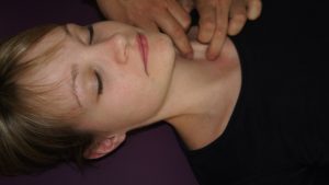 Fig. Technique Internal jugular vein
External jugular vein
Hand position: One middle finger is positioned posterior to the mandibular angulus on the vein. The other middle finger is positioned above the middle of the clavicle in the caudal area of the vein. execution: As for the technique for the internal jugular vein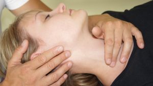 Fig. external jugular vein
Basilar plexus, marginal sinus
Hand position: The middle, index and ring fingers of both hands are positioned as close as possible to the foramen magnum from the posterior and lateral sides. execution: The fingers exert rhythmic pressure on the basilar plexus at the clivus and on the marginal sinus around the foramen magnum of the occiput. The aim is to support venous outflow from the superior and inferior petrosal sinuses and from the cavernous sinus into the basilar plexus and further into the marginal sinus.Fig. technique for the basilar plexus and marginal sinus
Equalization of the autonomic nervous system
This is achieved, for example, by establishing an osteopathic “felt sense” or through the technique of “heart-focused palpation” (see Liem 2017).
Ventricle
Compression of the 4th ventricle (CV-4): According to Magoun, this acts as a lymphatic pump (Magoun 1976) and should generally lead to an improved supply of cells, improved lymphatic movement and tissue regeneration as well as stimulation of the cranial nerve nuclei in the area of the 4th ventricle. Compression of the lateral ventricles and the 3rd ventricle are also possible (Liem 2013).
Drainage of the cisterns
Drainage techniques for the cisterns, e.g. the cisterna cerebellomedularis, can also be performed. To do this, the little fingers and/or ring fingers are placed below the inion and the thumbs are placed posterior to the porus acusticus externus. Rhythmic pressure is then applied to the cisterna cerebellomedullaris, i.e. the space between the cerebellum and the medulla. PHOTO Fig: Drainage of the cisterna cerebellomedullaris
Durale techniques
This includes, for example, the drainage technique of the superior sagittal sinus. These techniques are used with the aim of stimulating lymph drainage and LCS reflux into the dural sinus. To do this, the index finger and thumb are placed along the superior sagittal sinus on the frontal, parietal and occipital bones. The thumbs touch each other. Anteroposterior and craniocaudal rhythmic compression and decompression is performed on the dural sinus system. PHOTOFig: Falx technique for drainage of the superior sulcus sinusFor treatment of the transverse sinus, sigmoid sinus and superior petrosal sinus, the fingers can be placed along the base of the tentorium and the sinuses rhythmically drained (photo). For further dural techniques, see Liem (2013) Fig: Tentorial technique for drainage of the transverse, sigmoid and superior petrosal sinuses
OA release
To improve venolymphatic drainage from the head area, see Liem (2013).
Pumping technology at the Kranium
The Bjornas technique (personal communication between the author and Kjell Erling Bjornaes 2017) is used to stimulate lymph flow. Both hands embrace the skull, the thumbs lie on both sides along the sagittal sutura, the index and middle fingers anteriorly in front of the ear, the ring finger on the occipitomastoid sutura and the little finger on the occipital bone. Gentle, intermittent pressure is applied at a frequency of 10 to 12 times per minute for about three minutes. If necessary, the venous angles in the area of the clavicle and the 1st rib must be released.
Deep cervical lymph nodes and general lymph flow improvement
The lymphatic drainage techniques of the deep cervical lymph nodes and for general lymph flow improvement in the head area are used here to improve drainage, taking into account the LCS lymph connection (see above). Contraindications for lymphatic pumping techniques are
- untreated malignant tumor
- Acute inflammation with fever
- Thrombosis
- decompensated heart failure
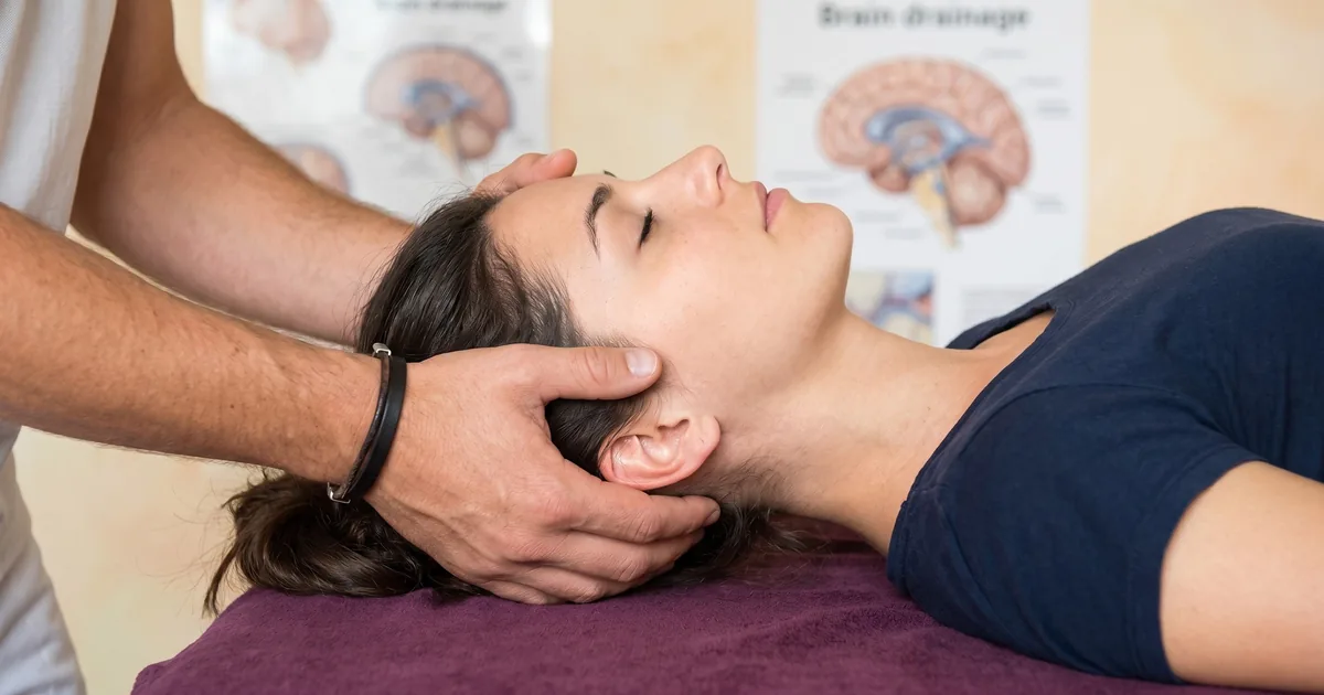
Drainage of the deep cervical lymphatic vessels: To drain the deep lymphatic vessels, the neck can be alternately compressed and decompressed in combination with lateral inclination and rotation movements in the neck. Repeat this five times (it is particularly important to repeat alternating movements in the neck area five times [personal communication of the author with Jean-Paul Belgrado 2017]).Fig: Drainage of the deep cervical lymph vessels
- Venolymphatic pump technique on the clavicle: A rhythmic pumping movement is exerted on the medial end of the clavicle while the head is stabilized in slight extension and contralaterally (Liem 2010).
General lymphatic flow improvement in the head area: The most efficient pressure to drain interstitial fluid into the initial lymphatic network is about 60-80 mmHg. This is more pressure than normally applied in lymphatic techniques (author’s personal communication with Jean-Paul Belgrado 2015). In a pilot study by Roth et al. (2016), craniocervical manual lymphatic drainage was used for the first time to reduce intracranial pressure in acute brain diseases.
- Place the fingers supraclavicularly on both sides. While the neck is stretched out, the fingers exert gentle pressure in the region of the lymphatic reflux. In addition, negative pressure can be created by making an inhaling movement with the chest without inhaling air.
- Then drain the lymph three times each on the face, the side of the head and the back of the head from cranial to caudal in the direction of the medial clavicle.
- Drain cranially from the navel towards the manubrium.
This sequence can also be shown to the patient as a self-help technique. Simply moving the skin is enough to increase the lymph flow by a factor of 20 (Ikomi et al. 1985).self-help technique to stimulate lymph flow: place the tongue on the roof of the mouth. After inhaling deeply and exhaling completely, make an inhaling movement with the chest without inhaling air. This creates negative pressure in the thorax and stimulates lymph flow. Lymphatic pump in the chest and abdominal area and on the feet: to stimulate the entire lymphatic system (see Liem 2013).
Drainage of the nose
The Chapman reflexes of the nose are also tested here: anterior: 2nd costal cartilage and 1st intercostal space, about 8 cm lateral to the sternum, posterior: midway between the processus spinosus and processus transversus of the axis (Liem 2010)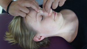 Fig: Chapman reflexes for the noseTreatmentof mouth breathing: For example, the root of the nose can be grasped with the index finger and thumb, while the nostrils are grasped with the other hand to induce rhythmic drainage.Rhythmic drainage technique in the area of the lamina cribrosa: Alternative techniques for the lamina cribrosa can be found, for example, in Liem (2010)Drainage technique for the olfactory nerve: First, a high cervical flexion and a contralateral lateral inclination of the cervical spine are set. In this presetting, the ethmoid bone in the area of the lamina cribrosa, the nasal septum and/or the conchae are rhythmically compressed and decompressed.
Drainage of the ear
Ear traction technique: Drainage by rhythmic traction on the auricle caudally, posteriorly or cranially. Drainage technique for the vestibulocochlear nerve: First, a high cervical flexion and a contralateral lateral tilt of the cervical spine is set. In this preset position, rhythmic compressions and decompressions are then performed on the temporal bone, especially on the mastoid or pars petrosa, occipital bone and sphenoid bone.
Cranial nerves
Drainage of cranial nerves IX, X, XI: First, a high cervical flexion and a contralateral lateral tilt of the cervical spine is set. The respective cranial nerve sheaths are then drained rhythmically on one side. Drainage of the upper cervical nerves and nerve sheaths: The index, middle and ring fingers are placed close to the vertebral foramina of the upper cervical nerves. While rhythmically flexing and extending the neck, a gentle homolateral cranial traction and a homolateral rotation to the side of the drainage are simultaneously applied (Liem 2017).Photo: Drainage of the upper cervical nerves and sheathsOther techniques: A decongestion of the pharynx and tonsils (Liem 2010) and technique of the internal carotid artery and internal carotid artery. According to Still, the fascial-lymphatic system could be stimulated by influencing the blood circulation via the nerves. (Still 1902). Correspondence address:Torsten LiemOsteopathie Schule DeutschlandWeidestraße 118 c22083 Hamburg
Literature
[1] Brinker T, Stopa E, Morrison J, Klinge P. A new look at cerebrospinal fluid circulation. Fluids Barriers CNS 2014; 11: 10[2] Engelhardt B, Carare RO, Bechmann I, Flüge A, Laman JD, Weller, RO. Vascular, glial, and lymphatic immune gateways of the central nervous system. Acta Neuropathol 2016; 132: 317-338[3] Ikomi F, Hunt J, Hanna G, Schmid-Schönbein GW. Interstitial fluid, plasma protein, colloid, and leukocyte uptake into initial lymphatics. J Appl Physiol 1985; 81 (5): 2060-7[4] Kiviniemi V, Wang X, Korhonen V, Keinanen T, Tuovinen T, Autio J, LeVan P, Keilholz S, Zang YF, Hennig J, Nedergaard M: Ultra-fast magnetic resonance encephalography of physiological brain activity – Glymphatic pulsation mechanisms? J Cereb Blood Flow Metab 2016; 36 (6): 1033-1045[5] Lehtinen P. Unraveling the link between brain and lymphatic system. Wihuri Research Institute 2016. http://www.wri.fi/unraveling-the-link-between-brain-and-lymphatic-system. Accessed: 22.6.2017[6] Liem T. Osteopathic treatment of the dura. In: Liem T, Tozzi P, Chila A (eds.) Fascia in the osteopathic field. Edinburgh: Handspring, 2017, p. 547f.[7] Liem T. Osteopathie: Ein praktisches Lehrbuch, 6th ed. Stuttgart: Haug, 2013[8] Liem T. Praxis der Kraniosakralen Osteopathie, 3rd ed. Stuttgart: Haug, 2010[9] Liem T. Treatment Principles. In: Liem T, Heede P (eds.) Foundations of morphodynamics in osteopathy. Edingburgh, Handspring, 2017, p. 353, 358[10] Louveau A, Smirnov I, Keyes TJ, Eccles JD, Rouhani SJ, Peske JD, Derecki NC, Castle D, Mandell JW, Lee KS, Harris TH, Kipnis J. Structural and functional features of central nervous system lymphatic vessels. Nature 2015; 523: 337-341[11] Magoun HI. Osteopathy in the cranial field, 3rd ed. Kirksville: Journal Printing Company, 1976, p. 110[12] Moser M, Liem T. Biological rhythms and their significance in osteopathy. In: Liem T, Heede P (eds.) Foundations of morphodynamics in osteopathy. Edingburgh: Handspring, 2017, p. 39-66[13] National Institutes of Health. Lymphatic vessels discovered in central nervous system. NIH Research Matters 2015. https://www.nih.gov/news-events/nih-research-matters/lymphatic-vessels-discovered-central-nervous-system. Accessed: 16.7.2015[14] Roth C, Stitz H, Roth C, Ferbert A, Deinsberger W, Pahl R, Engel H, Kleffmann J. Craniocervical manual lymphatic drainage and its impact on intracranial pressure – a pilot study. Eur J Neurol 2016; 23 (9): 1441-6[15] Sá-Pereira I, Brites D, Brito MA. Neurovascular unit: a focus on pericytes. Mol Neurobiol 2012; 45 (2): 327-347[16] Still AT. The philosophy and mechanican principles of osteopathy. Kansas City, Missouri: Hudson-Kimberly, 1902