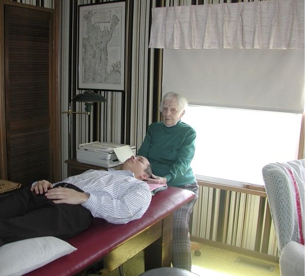
Anne Wales graduated from the Kansas City College of Osteopathy and Surgery and subsequently practised osteopathy for over 50 years in Rhode Island. She later lived in Massachusetts. She was also active in teaching. She served as editor of “Teachings in the Science of Osteopathy” by W.G. Sutherland and “Contributions of Thought. The Collected Writings of W.G. Sutherland” and is often regarded as the direct legacy of W.G. Sutherland. Anne Wales died on August 1, 2005, at the age of 101.The following interview, which was published as early as 2/2001 in the journal “Osteopathic Medicine”, is a compilation of personal conversations and correspondence between Anne Wales D.O. and Torsten Liem D.O. from 1996 to 2001.Especially in today’s fast-paced times, it seems incredible—and very humble—that a man would research and reflect for more than twenty years before sharing his results with the public. Can you tell us something about Sutherland’s career path and his contributions to osteopathy? Dr Sutherland was a journalist and editor of the Herald in Austin, Minnesota, when he first came into contact with osteopathy. He attended the American School of Osteopathy in Kirksville and graduated with the class of 1900. From 1900 to 1944, he practised osteopathy in Minnesota. He then began teaching about the human skull. He taught in California from 1944 until shortly before his death in September 1954. Over several decades, he studied the mechanical influences of the articular surfaces of the cranial and facial bones on the living human head. Sutherland’s great achievement was integrating the head into the osteopathic model, so that osteopathic treatment could address not only the feet but also the head and face. There is no longer just one answer to a patient’s presentation, but many treatment options tailored to the patient’s needs. Can you tell us something about Sutherland’s courses? We attended Dr Sutherland’s courses in Des Moines, Iowa, Chicago, Illinois, Providence, Rhode Island, and I learned a great deal in his teaching about the practice of osteopathy, as did many others. His teaching was based on the mechanics of the articular surfaces of the skeletal system, especially the movement of the cranial bones. He wanted his students to learn to see the anatomy of the articular surfaces in their mind’s eye. Dr Sutherland was a good teacher and an original thinker whose foundations were based on the anatomy of the human body. G. Sutherland said that the goal of osteopathic treatment was to create better exchange among all the body’s fluids across all contact surfaces. I understood that the human body is 70% water—half of it inside the cells. 35% is within the blood system. So how do air, water and food get into the cells?! Sutherland regarded the cranial cavity as a modified spherical space (the space in which the brain lies). If any joint within it moves, everything else moves as well. In this way, its shape changes. All cranial bones move in order to do this. You described some Sutherland techniques to me that differ significantly from Lippincott’s accounts published in Sutherland’s book “Teaching in the Science of Osteopathy”. From 1945 to 1950, my husband and I attended the Lippincott Cranial Study Group meetings in Moorestown, New Jersey. I have not read Lippincott’s written accounts. Howard A. Lippincott studied at the American School of Osteopathy in Kirksville, Missouri, with the class of 1918. Rebecca C. Lippincott was in the class of 1923 at the Philadelphia College of Osteopathy. In 1940, they studied with Dr Sutherland in Minnesota during their holidays. They wanted to pass on in writing what they themselves had learned. When you taught me the “point of balanced ligamentous tension”, it was more understandable to me than Lippincott’s descriptions. Could you briefly summarise again how you would describe a “Point of Balanced Ligamentous Tension” (PBLT) in the skeletal system, and when ligamentous dysfunctions can occur? The “point of balanced tension” in the skeletal system is where you position a joint in such a state of balance that the patient’s self-corrective forces move the bone into the correct position. Ligamentous dysfunctions can occur with strains, sprains, subluxations, dislocations and fractures. What other factors should be applied in the treatment of movement restrictions?Balanced ligamentous tension, approximation, traction, support through the patient’s breathing, support through the patient’s posture, active support, “fluid drive”, directing the “tide” (tidal motion). In which structures in the body do you establish a “point of balanced membranous tension”?In the cranium, in the forearm, and between the fibula and tibia. This is supported by techniques that direct the “tide” (tidal motion). You use the term “facial drag” more frequently. Could you explain it for us in a little more detail? In standing, with both feet on the ground and the arms hanging down at the sides, the centre of gravity is in the pelvis and the line of gravity runs through the dens of the axis. “Drag” means a sinking of tissues into the ventral region of the body. Which structures are particularly affected? The diaphragm and all attachments of the diaphragm. The colic flexures, the liver and spleen capsules, and the pericardium. The thorax is stabilised by the mediastinal fascia and the neck by the prevertebral fascia. Superiorly, the posterior neck and shoulder musculature attaches externally to the occipital bone. The anterior and posterior longitudinal ligaments connect the occipital bone to the cervical vertebrae. The prevertebral fascia attaches to the basilar part of the occipital bone directly behind the pharyngeal tubercle. The pharyngeal raphe is attached externally to the petrous part of the temporal bone and to the free margins of the medial pterygoid processes. This forms the nasopharynx. The perpendicular plate of the ethmoid and the vomer articulate with the body of the sphenoid… Altogether, there are many influences that can exert traction on the outer surface of the cranial base and the upper neck region.Inside the cranium, the squama is a special area. All dural duplications meet there. As taught by Dr Sutherland, the falx cerebri and the tentorium cerebelli—the inner layer of the dura mater—function as a reciprocal tension mechanism within the cranium. The dural duplications join in the straight sinus and house the venous sinus, which transports venous blood to the jugular foramina. According to Dr Sutherland, the inner layer of the dura mater moves the cranial bones at the sutures so that everything together can change its shape in a physiological rhythm. It acts as passive coordination of movement at the joint-like connections of the cranial bones. Even in the treatment of the facial bones, Dr Sutherland mentions a “point of balanced membranous tension”, although the dura has no direct contact there.Because the facial skeleton is connected to the cranial base, it is strongly influenced by the cranial base—even bones that have no direct connection to the cranial base, such as the maxilla. Here, the influence of the cranial base is transmitted via the articular surfaces from the sphenoid bone to the palatine bone and from there on to the maxilla. What are the influences on the movement of the cranial bones and the sacrum? The brain lies in a water bed—surrounded externally by cerebrospinal fluid in the subarachnoid space and embedded in the cisterns, and with cerebrospinal fluid internally in the ventricles of the brain—like a house in the ocean, a house with open doors. Due to the fluctuation of cerebrospinal fluid and the inherent motility of the brain tissue, the container (meaning the bony skull) continuously changes its shape, e.g. at the sphenobasilar synchondrosis. If the midline from the nasal septum to the coccyx flexes, all paired structures go into external rotation, and vice versa. At the foramen magnum, the inner layer of the dura mater is firmly attached—also to the posterior longitudinal ligament and to the upper cervical vertebrae. The dura then hangs relatively loosely in the spinal canal down to the second sacral vertebra. Thus, the sacrum is part of the inherent mechanism. Still already said that the nerves would drink the cerebrospinal fluid. The meninges follow the nerves. After leaving the brain, the cerebrospinal fluid becomes tissue fluid. The term changes, but there is a certain connection and continuity. Please explain the difference between the longitudinal “tide” (fluctuation) and the transverse “tide” (fluctuation)?In my view, the longitudinal fluctuation of cerebrospinal fluid (CSF) is a physiological phenomenon. The movement of the structures of the PRM in flexion and extension is related to the longitudinal fluctuation. When the fluctuation appears, I perceive during palpation as if the cisterns and the cerebellum were expanding, and the fluctuation continues along the falx. Lateral fluctuation is usually induced by a technique. At any time, I could place the thenar eminences of my hands on the mastoid processes, or place my hands on the greater wings of the sphenoid, the parietal bones or the sacrum, and induce a lateral fluctuation. This is a process for directing the “tide”. You can compress the fourth ventricle (CV-4) by changing the shape of the posterior cranial fossa beneath the tentorium cerebelli. For this, the hands must contact the supraocciput. This achieves a certain clinical effect—the same as when I induce an alternating lateral fluctuation. Once I was asked to treat Dr Sutherland. So I visited him at home. His wife opened the door and led me to Dr Sutherland. He was already lying down, and after I greeted him, I sat at the head of the table. I asked Dr Sutherland what I should do. He said I should place my hands on his head. He took my hands and placed them on the mastoid processes, with my fingers crossed under his neck. I said nothing. He said nothing. So I began. I followed what I felt. His head moved from one side to the other. After some time, he asked me whether I could feel anything. I said the head was moving from one side to the other. Very good, he said, now bring the movement to a stop. You do that by following the extension until it comes to a stop. And then you stay there, he said. You can do the same with internal and external rotation at the parietal bones. For example, if the patient is having an epileptic seizure and you want to stop it, gently bring the parietal bones or the sacrum into internal rotation/extension. Another time, I had a patient with a livid bluish, severely swollen hand. It was so painful that I could not touch it. I considered what I could do. I made a fist and asked the patient to grasp my fist with their hand. I induced a lateral fluctuation in the hand and a stop. And their skin colour changed. And then I noticed that we were breathing in synchrony. The swelling of the hand virtually disappeared. What do you think about the described axes of motion of the cranial bones? In my palpatory experience, I have often had the impression that the theory of axes of motion reflects only very imprecisely what is directly perceptible when my hands palpate the skull. And at the beginning, the biomechanical model describing cranial bone motion made it somewhat difficult for me to come into direct contact with the tissue without projecting that model into my palpatory experience. The bony skull is a three-dimensional container for the brain. There are sutures between the individual cranial bones. All these joint-like connections move in space. If a single bone moves, all the others must move at the same time. I do not necessarily think in terms of the so-called axes of motion of the individual cranial bones; I think more in terms of the different articular surfaces of the cranial bones. Everything has to move all at once. These days, there are many discussions about rhythms: Magoun (10–14/min.), Upledger (6–12/min.), Becker (6–10/10 min.), Jealous (2.5/min.). Did Sutherland himself ever state a frequency of the Primary Respiratory Mechanism? And what do you think about the different rhythms?I do not know of any specific frequency of the Primary Respiratory Mechanism, and I cannot remember Dr Sutherland counting the rhythm of the fluctuations. The Woods physicians introduced the term “Cranio Rhythmic Impulse” and counted the rhythm. In your view, what is special about an osteopath?As an osteopath, you examine the patient’s body with your hands. You study anatomy so that you can understand how the body works and what the problem is that brings the patient to you. You want to understand the issue before prescribing any kind of treatment. You want to understand what their complaints are, the history of their complaints, and then you want to find out what the problem behind their complaints is. Anne, how is it that you still look so young at your advanced age? Is it an osteopathic secret? I have received many treatments since I attended college in 1922. I began practising osteopathy in 1927 and ended my practice in 1977. I try to respect my physical limits and to move within them. Even today, I still treat my family, friends and close colleagues—that is, I give about one to three treatments a day.




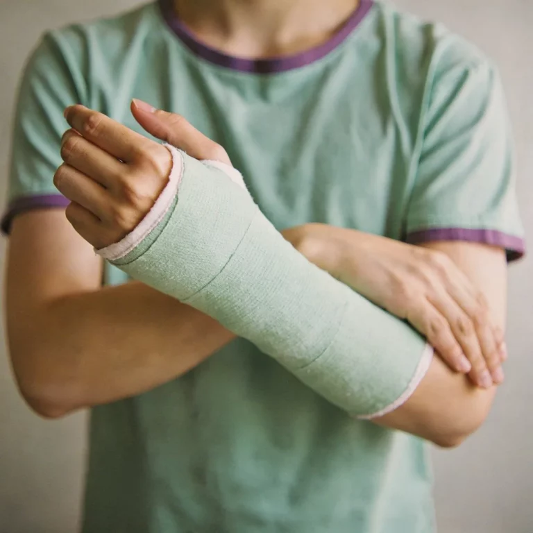
Radial Head Fracture: Osteopathic Treatment Step by Step
Torsten Liem, CEO of Osteopathie Schule Deutschland, is not only an expert in this field but also founded the Osteopathic Research Institute as well as